Friday, November 20, 2009
Monday, November 9, 2009
Brain Structures and their Functions
The nervous system is your body's decision and communication center. The central nervous system (CNS) is made of the brain and the spinal cord and the peripheral nervous system (PNS) is made of nerves. Together they control every part of your daily life, from breathing and blinking to helping you memorize facts for a test. Nerves reach from your brain to your face, ears, eyes, nose, and spinal cord... and from the spinal cord to the rest of your body. Sensory nerves gather information from the environment, send that info to the spinal cord, which then speed the message to the brain. The brain then makes sense of that message and fires off a response. Motor neurons deliver the instructions from the brain to the rest of your body. The spinal cord, made of a bundle of nerves running up and down the spine, is similar to a superhighway, speeding messages to and from the brain at every second.
The brain is made of three main parts: the forebrain, midbrain, and hindbrain. The forebrain consists of the cerebrum, thalamus, and hypothalamus (part of the limbic system). The midbrain consists of the tectum and tegmentum. The hindbrain is made of the cerebellum, pons and medulla. Often the midbrain, pons, and medulla are referred to together as the brainstem.
The Cerebrum:The cerebrum or cortex is the largest part of the human brain, associated with higher brain function such as thought and action. The cerebral cortex is divided into four sections, called "lobes": the frontal lobe, parietal lobe, occipital lobe, and temporal lobe. Here is a visual representation of the cortex:
What do each of these lobes do?
• Frontal Lobe- associated with reasoning, planning, parts of speech, movement, emotions, and problem solving
• Parietal Lobe- associated with movement, orientation, recognition, perception of stimuli
• Occipital Lobe- associated with visual processing
• Temporal Lobe- associated with perception and recognition of auditory stimuli, memory, and speech
Note that the cerebral cortex is highly wrinkled. Essentially this makes the brain more efficient, because it can increase the surface area of the brain and the amount of neurons within it. We will discuss the relevance of the degree of cortical folding (or gyrencephalization) later.
A deep furrow divides the cerebrum into two halves, known as the left and right hemispheres. The two hemispheres look mostly symmetrical yet it has been shown that each side functions slightly different than the other. Sometimes the right hemisphere is associated with creativity and the left hemispheres is associated with logic abilities. The corpus callosum is a bundle of axons which connects these two hemispheres.
Nerve cells make up the gray surface of the cerebrum which is a little thicker than your thumb. White nerve fibers underneath carry signals between the nerve cells and other parts of the brain and body.
The neocortex occupies the bulk of the cerebrum. This is a six-layered structure of the cerebral cortex which is only found in mammals. It is thought that the neocortex is a recently evolved structure, and is associated with "higher" information processing by more fully evolved animals (such as humans, primates, dolphins, etc). For more information about the neocortex, click here.
The Cerebellum: The cerebellum, or "little brain", is similar to the cerebrum in that it has two hemispheres and has a highly folded surface or cortex. This structure is associated with regulation and coordination of movement, posture, and balance.
The cerebellum is assumed to be much older than the cerebrum, evolutionarily. What do I mean by this? In other words, animals which scientists assume to have evolved prior to humans, for example reptiles, do have developed cerebellums. However, reptiles do not have neocortex. Go here for more discussion of the neocortex or go to the following web site for a more detailed look at evolution of brain structures and intelligence: "Ask the Experts": Evolution and Intelligence
Limbic System: The limbic system, often referred to as the "emotional brain", is found buried within the cerebrum. Like the cerebellum, evolutionarily the structure is rather old.
This system contains the thalamus, hypothalamus, amygdala, and hippocampus. Here is a visual representation of this system, from a midsagittal view of the human brain:
Thalamus
Thalamus- a large mass of gray matter deeply situated in the forebrain at the topmost portion of the diencephalon. The structure has sensory and motor functions. Almost all sensory information enters this structure where neurons send that information to the overlying cortex. Axons from every sensory system (except olfaction) synapse here as the last relay site before the information reaches the cerebral cortex.
Hypothalamus
Hypothalamus- part of the diencephalon, ventral to the thalamus. The structure is involved in functions including homeostasis, emotion, thirst, hunger, circadian rhythms, and control of the autonomic nervous system. In addition, it controls the pituitary.
a coronal view
Amygdala
Amygdala- part of the telencephalon, located in the temporal lobe; involved in memory, emotion, and fear. The amygdala is both large and just beneath the surface of the front, medial part of the temporal lobe where it causes the bulge on the surface called the uncus. This is a component of the limbic system.
Hippocampus
Hippocampus- the portion of the cerebral hemisphers in basal medial part of the temporal lobe. This part of the brain is important for learning and memory . . . for converting short term memory to more permanent memory, and for recalling spatial relationships in the world about us
Brain Stem: Underneath the limbic system is the brain stem. This structure is responsible for basic vital life functions such as breathing, heartbeat, and blood pressure. Scientists say that this is the "simplest" part of human brains because animals' entire brains, such as reptiles (who appear early on the evolutionary scale) resemble our brain stem. The brain stem is made of the midbrain, pons, and medulla. Click on the words to learn what these structures do:
Midbrain/Mesencephalon
Midbrain/ Mesencephalon- the rostral part of the brain stem, which includes the tectum and tegmentum. It is involved in functions such as vision, hearing, eyemovement, and body movement. The anterior part has the cerebral peduncle, which is a huge bundle of axons traveling from the cerebral cortex through the brain stem and these fibers (along with other structures) are important for voluntary motor function.
Pons
Pons- part of the metencephalon in the hindbrain. It is involved in motor control and sensory analysis... for example, information from the ear first enters the brain in the pons. It has parts that are important for the level of consciousness and for sleep. Some structures within the pons are linked to the cerebellum, thus are involved in movement and posture.
Medulla
Medulla Oblongata- this structure is the caudal-most part of the brain stem, between the pons and spinal cord. It is responsible for maintaining vital body functions, such as breathing and heartrate
note: There will be illustrations to be given in class
Reference: http://serendip.brynmawr.edu/bb/kinser/Structure1.html
Thursday, October 1, 2009
The terms
Definition of terms
1. Origin- also called the head , the most stationary end of muscle
2. Insertion- the end of the muscle attached to the bone undergoing the greatest movement.
3. Agonist- a muscle that accomplishes a certain movement.
4. Antagonist- A muscle acting on opposition.
5. Tendon- a tough connective tissue band connecting muscles to bone.
6. Abduction-movement away from the median or midsagittal plane
7. Adduction- movement toward the median
8. Pronation-rotation of the forearm so that the palm is down
9. Supination- The palms face up
10. Flexion- moves part of the body in the anterior or ventral to the coronal plane
11. Extension- Moves a part in a posterior or dorsal coronal plane
12. Protraction- movement in which structure, such as mandible, glides anteriorly
13. Retraction- the structure glides posteriorly.
14. Excursion -the movement of the structure to one side or other.
Reference: Seeley, Stephens et al. Essentials of Anatomy and Physiology 6th edition, McGraw-Hill International Edition.
Tuesday, September 22, 2009
The Skeletal Muscle
Objectives
At the end of the 60-minute period, at least 85% of the learners are expected to:
a. describe the structure and characteristics of skeletal muscles through small group discussion,
b. appreciate the role played and the characteristics of skeletal muscles
c. list down the different structures of the skeletal muscles.
Reference: SeeleyR.R., et al Essentials of Anatomy and Physiology 6th Edition, Mc-Graw Hill International.
Topic: Functions of the Muscles
Characteristics of Skeletal Muscles
Structure of Skeletal Muscles
I. Functions of the Skeletal Muscles
a. Body Movement. Contraction of the skeletal muscle is responsible for the overall movement of the body such as walking, running etc.
b. Maintenance of posture. Skeletal muscles constantly maintain tone, which keeps us sitting or standing erect.
c. Respiration. Muscles of the thorax are responsible for movement necessary for breathing.
d. Production of body heat. When skeletal muscles contract, heat is given off as a by-product.
e. Communication. Skeletal muscles are involved in all aspects of communication.
f. Constriction of organs and vessels.
g. Heart beat
II. Characteristics of Skeletal muscles
a. Contractility is the ability of skeletal muscles to shorten with force.
b. Excitability is the capacity of skeletal muscles to respond to stimuli.
c. Extensibility means that skeletal muscles can be stretched.
d. Elasticity is the ability of the skeletal muscles to recoil to their original resting length after they have been stretched.
III. Structure
Each skeletal muscle is surrounded by a connective tissue sheath called EPIMYSIUM or FASCIA. A muscle is composed of numerous visible bundles called MUSCLE FASCICULI which are surrounded by loose connective tissue called the PERIMYSIUM. A fasciculus is composed of several muscle cells or muscle fibers. Each muscle fiber is surrounded by loose connective tissue called ENDOMYSIUM.
The muscle contraction is much easier to understand when we understand the structure of a muscle cell. The cytoplasm of each of the muscle fiber is called the sarcoplasm, contains numerous myofibrils. Each myofibril is a thread like structure that extends from one end of the muscle fiber from one end of the muscle fiber to another. Myofibril contains two major kinds of protein fibers: myosin and actin.
The actin and myosin myofilaments are arranged into highly ordered repeating units along with the myofibril called sarcomere. Sarcomere is the basic structural units of the skeletal muscle. Each sarcomere extends from one Z disc to another Z disc.
Actin filaments or the thin filaments, resemble two-minute strands of pearl twisted together. Troponin molecules are attached at specific intervals along with actin myofilaments and provide calcium binding sites on the actin myofilaments. Tropomyosin filaments are located along the groove between the twisted strands action myofilaments subunits.
Myosin filaments of thick filaments, resembles bundles of minute golf clubs. The part of the myosin molecule that resembles the golf club heads can bind to the exposed attachment sites on the actin myofilaments.
The cell membrane of the muscle fiber is called the sarcolemma. The T-tubles are associated with a highly organized Smooth endoplasmic Reticulum called the Sarcoplasmic reticulum. It has a high concentration of calcium ion which is responsible for the muscle contraction.
The arrangement of the actin and myosin myofilaments in sarcomeres gives the myofibril a banded appearance. The light I band, which consists of only myosin myofilament, spans each Z disk and ends at the myosin myofilaments. A darker, central region in each sarcomere, called an A band extends the length of the myosin myofilaments. In the cenetrr of each filament is a second light zone called the H zone which consists of only myosin myofilament. The myosin myofilaments are anchored in the center of the sarcomere at a dark-staining band, called the M-line.

Skeletal Muscle
Sunday, September 20, 2009
Saturday, July 25, 2009
Vertebrate Integumentary System
THE INTEGUMENTARY SYSTEM
The Vertebrate’s outer covering….i.e. the skin
FUNCTIONS:
1. Support and protection (primary function)
2. Reception/transduction of ext. stimuli
3. Material transport (excretion, resorption, dehydration, rehydration)
4. Thermoregulation
5. Gas exchange
6. Nutrient storage
7. Locomotion
8. Behavior (sexual selection, aggression, identification)
9. Sound production
SKIN IS FUNCTIONALLY A UNIT WITH 3 PARTS:
1. Epidermis
2. Dermis
3. Basement Membrane Complex
The Epidermis:
Outermost layer
An Ectodermal derivative
Often glandular
The Dermis (=corium)
Innermost layer
A mesodermal derivative
Neural Crest gives rise to chromatophores or dermal
armor if present
Contains a neural and vascular supply
The Basement Membrane Complex
In between epidermis and dermis
Outer single layer = Basal Lamina
Inner layers = Basal Lamella
SURVEY OF VERTBRATE SKIN
Amphioxus
Epidermis limited to columnar cells and mucous cuticle
Agnatha
Epidermis is more complex with club and granule cells
The Fishes
General characteristics:
Epidermis is very thin, with 2 cell types….epidermal
cells and unicellular glands (mucous)
Mucous cuticle on surface
Microridges to hold mucous in place
Dermis contains chromatophores
Three types of chromatophores:
Melanophores (brown or black pigment)
Lipophores (xanthophores with yellow pigment and
erythrophores with red pigment)
Iridophores (reflective)
Dermis produces a dermal scale in many
Chondrichthyes:
Placoid scales or dermal denticles
Outer enamel; inner dentin
Epidermis does not cover scales
Osteichthyes:
Bony fish scales are covered by a thin layer of epidermis
Osteichthyes-Sarcopterygii:
Cosmoid Scales
Dermally derived
Outer enamel, intermediate dentin, bony core
Osteichthyes-Actinopterygii:
Dermal scales of three basic types….
Ganoid (Gars, Bichirs)
Dermally derived
Outer enamel (=ganoin), inner bone
Cycloid and Ctenoid (Teleosts that bear scales)
Dermally derived
Scales entirely of lamellar bone
Annuli and Circuli
Amphibia
Epidermis with thin stratum corneum and very little
keratin; Leydig cells
Dermis with chromatophores, poison glands and mucous
glands
Scales are rare
Reptilia
Epidermal scales, with thick outer layer of keratin
Thinner “hinge” region
Inner layer of epidermis regenerative….sloughing
Outer scale surface (Oberhäutchen) often sculpted
…microepidermatoglyphics
Dermis with chromatophores in many
Dermis may possess Osteoderms
Birds
Epidermis thin and bilayered…stratum corneum and
stratum basale
Dermis well-vascularized and innervated
Very few glands
Unique epidermal feathers (of keratin) with basic
structure:
Calamus (quill)
Rachis (shaft)
Barbs, barbules and hooklets
Basic feather types:
Flight, Down, Filoplume and Contour
Feathers probably arose as epidermal scale modifications
Epidermal chromatophores produce pigments which are
carried into feather during development, but
feather surface provides structural color
Feather development
Epidermal feather primordium, dermal papilla,
“collar” and eruption
Feathers grow in tracts, and are connected together in the
dermis by tiny feather muscles
Mammals
Epidermis with 5 layers:
Stratum corneum – outer, keratinized
Stratum lucidum – no organelles
Stratum granulosum – keratin development
Stratum spinosum – developing cells
Stratum basale – germination layer
Epidermal glands present in dermis:
Sebaceous (oil) - Holocrine
Sudoriferous (sweat) – Merocrine
Gland types based on fate of product:
Exocrine – ducted; product into ducts
Endocrine – ductless; product into blood
Gland types based on cellular mode of secretion:
Cytogenic – whole cells; testes and ovaries
Holocrine – product is entire cell contents
Sebaceous
Merocrine – product moves through cell membrane
often by exocytosis; salivary, pancreas
Apocrine – product is cytoplasm at tip of cell;
Mammary
Dermis well-vascularized and innervated
Hair produced in epidermis, and unlike scales and feathers
is an ingrowth of epidermis into the dermis
Root and Shaft
Cuticle, Cortex and Medulla
Fur (pelage) is a thick covering of hair
Guard hairs – longer, coarser
Underfur – shorter, finer
Hairs moved by arector pili muscles
Integumental Derivatives
Integumental derivatives result from on of three processes:
I. Functional Epithelial Extinction (FEE) which leads to “Structured Ectodermal Derivatives”
II. Ectodermal-Mesodermal Interaction (EMI)
which leads to “Structured Ectodermal-
Mesodermal Derivatives”
III. Delamination (DEL) which leads to “Structured
Mesodermal Derivatives”
I. Structured Ectodermal Derivatives
A. Integumental glands
Mucous – Fishes (unicellular)
Amphibians (multicellular)
Poison – Fishes (unicellular/multicellular)
Amphibian, one bird (multicellular)
Venom – Reptiles (modified salivary)
Platypus (modified sweat?)
Salivary – Primarily tetrapods
Musk (scent) – Reptiles, Mammals
Preen (uropygial) – Birds
Sebaceous(oil) - Mammals
Ceruminous (wax) – Mammals, Turkey
Sudorifereous (sweat) – Mammals
Mammary – Mammals (modified sebaceous?)
Photophore Glands – Deep sea fishes
B. Keratinized integument
A manifestation of FEE….several processes:
1. Shedding: Continuous loss of small flakes or cell groups. Probably in all vertebrates…even in areas of specialized thickenings (callouses, palmar and plantar surfaces).
2. Sloughing: Periodic loss of large complete sheets of skin.
Many fishes (mucous cuticle)
Most amphibians (with autophagy)
Reptiles (may be accompanied by autophagy)
Birds (feet)
Some seals, whales, elephants, cervid velvet
3. Molting: Periodic loss of specialized keratinized
ectodermal derivatives
Hair…..including baleen, quills
Feathers
Shell-breaker (=egg caruncle)…epidermal
structure of birds, turtles, crocodilians, tuatara
(Egg Tooth of lizards and snakes is a true tooth)
Turtle scutes
Lamprey “teeth”
Nuptial Pads
4. Retention: Rather permanent specialized
keratinized ectodermal derivatives.
Rattlesnake rattle
Beaks
Horn
True horn: bony spike from skull sheathed
in keratinized epidermis…never “shed”,
never branched (except for Pronghorn)
Not to be confused with:
Antlers (Usually branched,
no keratinization; often shed)
Giraffe “horns” (Bony core covered by
permanent epidermis..no
keratinization…never sloughed or “shed”)
Rhinoceros “horn” (fused mass of epidermal
hair-like papillae…no bone involved
Claws, Nails, Hoofs
All containing Unguis and Subunguis
Digital caps (amphibian “claws”)
Local thickenings – Tori, friction ridges
II. Structured Ectodermal-Mesodermal Derivatives
Composite structures derived from an interaction between Ectoderm and Mesoderm, such as
Dermal Scales
Teeth
III. Structured Mesodermal Derivatives
Structures derived primarily from Mesoderm, such as
Dermal Plates, or “Armor”
Armadillo
Crocodilian osteoderms
Turtle bony plates
Fat storage structure
Panniculus adiposus
Integumentary muscle
Panniculus carnosus
Bone
Reference:http://www.cst.cmich.edu/users/gilli1jc/Part%202%20Integument.htm
Tuesday, July 21, 2009
Tuesday, July 14, 2009
Monday, July 6, 2009
Protein
What Is Protein?
A protein is a long train of amino acids linked together. Proteins have different functions; they can provide structure (ligaments, fingernails, hair), help in digestion (stomach enzymes), aid in movement (muscles), and play a part in our ability to see (the lens of our eyes is pure crytalline protein).
Protein is a long chain molecule made up of amino acids joined by peptide bonds. Protein forms the structural material of bodily tissues.
Proteins, the principal constituents of the protoplasm of all cells, are of high molecular weight and consist essentially of combinations of a amino acids in peptide linkages.
Twenty different amino acids are commonly found in proteins and each protein has a unique, genetically defined amino acid sequence which determines its specific shape and function.
They serve as enzymes, structural elements, hormones, immunoglobulins, etc. And are involved in oxygen transport, muscle contraction, electron transport and other activities throughout the body and in photosynthesis.
Origin: Gr. Protos = first
The most important function of protein is to build up, keep up, and replace the tissues in your body. Your muscles, your organs, and some of your hormones are made up mostly of protein.
Protein also makes antibodies and hemoglobin (responsible for delivering oxygen to your blood cells).
Our body is able to produce 14 of the 20 amino acids. We have to get the remaining amino acids from the foods we eat.
fish. But proteins are complex and have many different functions within the human body.
Proteins form the major components of muscles, skin, tendons, blood vessels, hair, and cores of bones and teeth. They help you grow, heal wounds, and make up collagen - the connective tissue that gives your body its shape.
Other proteins, called enzymes, help generate energy in your body. Proteins called hormones act as internal “project managers”, ensuring your body runs itself properly. Insulin and glucagons, for example, are the hormones that control blood sugar levels. And proteins called antibodies are important components of your immune system, warding off foreign particles like bacteria.
Without protein, life would be impossible. Practically every cell in your body spends considerable time and energy manufacturing various kinds of proteins. Every imaginable part and function of your body has protein involved in some way, from the enzymes that are critical to the digestion of foods to the fibers that plug leaky blood vessels. But before you get carried away with putting protein on a nutritional pedestal, it can also be said that life would be impossible without sugars (carbohydrates) and fats (lipids).
Protein is simply a name for a nutritional family. We could just as easily have said this is a photo of the Jones or Smith family. To pick out their distinctive family characteristics we need to look closer. Do they all have large noses, blue eyes, and freckles? Just as human families have one trait in common with all othersthey are all members of the human speciesso do proteins. The principal trait for all proteins, large or small, is that they are composed of amino acids.
With about 20 different kinds available, every cell of your body can choose from the pool of amino acids to construct specific proteins to meet very diverse needs of human physiology. For example, there are boats to shuttle oxygen around (hemoglobin), taxis for small fats (LDL), and ballistic missiles to destroy invaders (immunoglobulins). Put two amino acids together and you get a dipeptide (di = "two"); put three amino acids together and the result is a tripeptide (tri = "three"); finally, a complex construction of many amino acids makes a polypeptide (poly = "many"). Human insulin, for instance, which is responsible for getting glucose into the muscles, is composed of 51 amino acids in two short polypeptide chains.
As the digestive process kicks into high gear, "intelligent" receptors on the large intestinal surfaces have to decide which amino acid, dipeptide, or tripeptide to absorb, and which will have to wait. This competition among the amino acids for absorption is not a big deal, since the body's receptors "know" which amino acids are needed most of the time. However, if your intestinal tract is flooded with amino acid supplements, the absorptive cells cannot deal with these refined and readily available amino acids all at once. Inevitably, the "pushier" amino acids, or the ones in greatest concentration, leave the receptors no choice but to take them in first.
The constant construction of new proteins, along with the destruction and elimination of old proteins, means that a continual supply of amino acids must be made available via nutrition to maintain a healthy balance. Different foods provide essential amino acids in varying proportions.
We now have clear evidence that even if a diet is derived exclusively from the vegetable kingdom, it can provide all the necessary amino acids for optimal health. It is generally sufficient simply to have a consistent supply of a variety of vegetables, legumes, and grains throughout the week.
Protein is one of the three macronutrients commonly identified as a dietary requirement. It represents nitrogen-containing compounds for which amino acids are the basic structural units.
Amino acids are small organic compounds containing at least one amino group and an organic acid group. The differences between amino acids lies in the differences between the amino acid side proteins are the most abundant organic compound of the body. More than fat, usually. Much more than carbohydrate. About 65% of the total body protein lies in the skeletal muscles.
Proteins function primarily in the growth and repair of body tissue. Just about every cell in our body has a protein component, and we are unable to synthesize new cells without the requisite building blocks. Hair, nails, skin contain protein. Blood contains plasma proteins; hemoglobin has a protein component. Proteins are components of some antibodies. Many hormones are proteins (like insulin). In fact, the protein content of the average cell is 16% of its total mass.
There are more than 50,000 different proteins in our bodies. These are all made from about 22 different amino acids. Our bodies can synthesize 14 of these 22 amino acids, we cannot make 8 of them, and these 8 must come from food. These 8 are called the essential amino acids. Sometimes we cannot synthesize other amino acids and therefore they too must come from diet.
Protein is a large, complex molecule composed of amino acids. The sequence of the amino acids, and thus the function of the protein, is determined by the sequence of the base pairs in the gene that encodes it. Proteins are essential to the structure, function, and regulation of the body. Examples are hormones, enzymes, and antibodies. Many bodybuilders use Protein to help build muscle in the body.
A protein is a complex, high molecular weight organic compound that consists of amino acids joined by peptide bonds. Proteins are essential to the structure and function of all living cells and viruses. Many proteins are enzymes or subunits of enzymes. Other proteins play structural or mechanical roles, such as those that form the struts and joints of the cytoskeleton. Still more functions filled by proteins include immune response and the storage and transport of various ligands. In nutrition, proteins serve as the source of amino acids for organisms that do not synthesize those amino acids natively.
Proteins are one of the classes of bio-macromolecules, alongside polysaccharides and nucleic acids, that make up the primary constituents of living things. They are amongst the most actively studied molecule in biochemistry and were discovered by Jöns Jacob Berzelius, in 1838.
Proteins are amino acid chains, made up from 20 different L-α-amino acids, also referred to as residues, that fold into unique three-dimensional protein structures. The shape into a which a protein naturally folds is known as its native state, which is determined by its sequence of amino acids. Below about 40 residues the term peptide is frequently used. A certain number of residues is necessary to perform a particular biochemical function, and around 40-50 residues appears to be the lower limit for a functional domain size. Protein sizes range from this lower limit to several hundred residues in multi-functional proteins. Very large aggregates can be formed from protein subunits, for example many thousand actin molecules assemble into a an actin filament. Large protein complexes with RNA are found in the ribosome particles, which are in fact 'ribozymes'.
Biochemists refer to four distinct aspects of a protein's structure:
Primary structure: the amino acid sequence Secondary structure: highly patterned sub-structures--alpha helix and beta sheet--or segments of chain that assume no stable shape. Secondary structures are locally defined, meaning that there can be many different secondary motifs present in one single protein molecule Tertiary structure: the overall shape of a single protein molecule; the spatial relationship of the secondary structural motifs to one another Quaternary structure: the shape or structure that results from the union of more than one protein molecule, usually called subunit proteins subunits in this context, which function as part of the larger assembly or protein complex. In addition to these levels of structure, proteins may shift between several similar structures in performing of their biological function. In the context of these functional rearrangements, these tertiary or quaternary structures are usually referred to as "conformations," and transitions between them are called conformational changes.
The primary structure is held together by covalent peptide bonds, which are made during the process of translation. The secondary structures are held together by hydrogen bonds. The tertiary structure is held together primarily by hydrophobic interactions but hydrogen bonds, ionic interactions, and disulfide bonds are usually involved too.
The two ends of the amino acid chain are referred to as the carboxy terminus (C-terminus) and the amino terminus (N-terminus) based on the nature of the free group on each extremity.
Two amino acids are combined in a condensation reaction. Notice that the peptide bond is in fact planar due to the delocalization of the electrons. The sequence of the different amino acids is considered the primary structure of the peptide or protein. Counting of residues always starts at the N-terminal end (NH2-group).
Proteins are involved in practically every function performed by a cell, including regulation of cellular functions such as signal transduction and metabolism. For example, protein catabolism requires only a few enzymes termed proteases.
Various molecules and ions are able to bind to specific sites on proteins. These sites are called binding sites. They exhibit chemical specificity. The particle that binds is called a ligand. The strength of ligand-protein binding is a property of the binding site known as affinity.
Since proteins are involved in practically every function performed by a cell, the mechanisms for controlling these functions therefore depend on controlling protein activity. Regulation can involve a protein's shape or concentration. Some forms of regulation include:
Allosteric modulation: When the binding of a ligand at one site on a protein affects the binding of ligand at another site. Covalent modulation: When the covalent modification of a protein affects the binding of a ligand or some other aspect of a the protein's function.
Proteins are generally large molecules, having molecular masses of up to 3,000,000 (the muscle protein titin has a single amino acid chain 27,000 subunits long). Such long chains of amino acids are almost universally referred to as proteins, but shorter strings of amino acids are referred to as "polypeptides," "peptides" or very rarely "oligopeptides". The dividing line is somewhat undefined, although a polypeptide may be less likely to have tertiary structure and may be more likely to act as a hormone (like insulin) rather than as an enzyme or structural element.
Reference: http://www.bionewsonline.com/5/what_is_protein.htm
Sunday, July 5, 2009
Tuesday, June 9, 2009
CHARACTERISTICS OF LIVING ORGANISMS
Cell
All living things are composed of one or more cells. Different types of cells have different "jobs" within the organism. Each life form begins from one cell, which then will split. These cells split, and so on. After this has happened several times, differentiation is undergone, when the cells change so that they are not the same thing anymore. Then they are used to begin to put together the final organism, some cells, for example, as the eyes, some as the heart, etc. The only arguable exception to this is viruses. They are not composed of cells, but are said to be "living."
Organization
Complex organization patterns are found in all living organisms. They arrange themselves on very small levels, grouping like things together. On larger levels, they become visible. This also has to do with differentiation, as the cells are organized in a manner that makes sense for the organism after they change to what they’ll be in the final organism.
Energy Use

All organisms use energy. The sum of the chemical energy they use is called metabolism. This energy is used to carry out everything they do. Autotrophs (plants) use energy from the sun for photosynthesis, to make their own ‘food’ (glucose). Heterotrophs (animals and humans) must ingest food for this purpose.
Homeostasis
All organisms have stable internal conditions which must be maintained in order to remain alive. These include temperature, water content, heartbeat, and other such things. In a way, this has to do with energy use, because a certain level of energy must be kept within the body at all times. For this, obviously, humans must then ingest food on a regular basis. Not all conditions are for the body to maintain itself; though most are.
Growth

All organisms grow and change. Cells divide to form new, identical cells. Differentiation happens, as well, when cells mutate into other types of cells, making a more complex organism. Organisms growing, changing, and becoming more complex is called development. Single-celled organisms do grow as well, but they will only become slightly larger – this is nearly unmeasurable.
Reproduction

All organisms reproduce in order to continue the species' life. This is combining genetic information (in sexual reproduction) or splitting into two organisms (in asexual reproduction) in order to create another of the same species. In sexual reproduction, the new organism will have some characteristics from the mother, and some from father. It may look like either of them, or it may not. In asexual reproduction, the new organism is an exact copy of the first. Sometimes, not every member of a species is able to reproduce. As long as others are (which we know they can, if they still exist today) then it does not threaten the species. (Except for mules, but don't worry about them, they are a bizarre anomoly.)
Response to stimuli:

A response can take many forms, from the contraction of a unicellular organism to external chemicals, to complex reactions involving all the senses of higher animals. A response is often expressed by motion, for example, the leaves of a plant turning toward the sun (phototropism) and by chemotaxis.
References:http://www.essortment.com/all/characteristics_rbrc.htm
http://en.wikipedia.org/wiki/Characteristics_of_living_things

All living things are composed of one or more cells. Different types of cells have different "jobs" within the organism. Each life form begins from one cell, which then will split. These cells split, and so on. After this has happened several times, differentiation is undergone, when the cells change so that they are not the same thing anymore. Then they are used to begin to put together the final organism, some cells, for example, as the eyes, some as the heart, etc. The only arguable exception to this is viruses. They are not composed of cells, but are said to be "living."
Organization

Complex organization patterns are found in all living organisms. They arrange themselves on very small levels, grouping like things together. On larger levels, they become visible. This also has to do with differentiation, as the cells are organized in a manner that makes sense for the organism after they change to what they’ll be in the final organism.
Energy Use

All organisms use energy. The sum of the chemical energy they use is called metabolism. This energy is used to carry out everything they do. Autotrophs (plants) use energy from the sun for photosynthesis, to make their own ‘food’ (glucose). Heterotrophs (animals and humans) must ingest food for this purpose.
Homeostasis

All organisms have stable internal conditions which must be maintained in order to remain alive. These include temperature, water content, heartbeat, and other such things. In a way, this has to do with energy use, because a certain level of energy must be kept within the body at all times. For this, obviously, humans must then ingest food on a regular basis. Not all conditions are for the body to maintain itself; though most are.
Growth

All organisms grow and change. Cells divide to form new, identical cells. Differentiation happens, as well, when cells mutate into other types of cells, making a more complex organism. Organisms growing, changing, and becoming more complex is called development. Single-celled organisms do grow as well, but they will only become slightly larger – this is nearly unmeasurable.
Reproduction

All organisms reproduce in order to continue the species' life. This is combining genetic information (in sexual reproduction) or splitting into two organisms (in asexual reproduction) in order to create another of the same species. In sexual reproduction, the new organism will have some characteristics from the mother, and some from father. It may look like either of them, or it may not. In asexual reproduction, the new organism is an exact copy of the first. Sometimes, not every member of a species is able to reproduce. As long as others are (which we know they can, if they still exist today) then it does not threaten the species. (Except for mules, but don't worry about them, they are a bizarre anomoly.)
Response to stimuli:

A response can take many forms, from the contraction of a unicellular organism to external chemicals, to complex reactions involving all the senses of higher animals. A response is often expressed by motion, for example, the leaves of a plant turning toward the sun (phototropism) and by chemotaxis.
References:http://www.essortment.com/all/characteristics_rbrc.htm
http://en.wikipedia.org/wiki/Characteristics_of_living_things
Monday, June 8, 2009
Origin of Life
SPECIAL CREATION THEORY

Creationism, Special creation is a theological doctrine which asserts that the origin of the universe and all life in it suddenly sprang into being by unconditional fiat or divine decree. Roman Catholicism has defined "special creation" in a different way. In Catholic teaching, the doctrine of immediate or special creation refers to the origin of the human soul.
In the creationist use of the phrase, special creation adheres to a literal interpretation of Genesis creation, accepting it as an accurate historical account of creation of the universe in its present form over the course of six twenty-four hour days. Special creation was the dominant theory of life's origins in the western world from the 16th century until the middle of the 19th century when it was supplanted by evolutionary thought.[1][2]
Duane Gish of the Institute for Creation Research defined "special creation" as being creation using supernatural processes:
We do not know how the Creator created, [or] what processes He used, for He used processes which are not now operating anywhere in the natural universe. This is why we refer to creation as special creation. We cannot discover by scientific investigation anything about the creative processes used by the Creator.
SPONTANEOUS GENERATION
or Equivocal generation is an obsolete theory regarding the origin of life from inanimate matter, which held that this process was a commonplace and everyday occurrence, as distinguished from Univocal generation, or reproduction from parent(s). The theory was synthesized by Aristotle[1], who compiled and expanded the work of prior natural philosophers and the various ancient explanations of the appearance of organisms; it held sway for two millennia. It is generally accepted to have been ultimately disproven in the 19th Century by the experiments of Louis Pasteur, expanding upon the experiments of other scientists before him (such as Francesco Redi who had performed similar experiments in the 17th century). Ultimately, it was succeeded by germ theory and cell theory.
The disproof of ongoing spontaneous generation is no longer controversial, now that the life cycles of various life forms have been well documented. However, the question of abiogenesis, how living things originally arose from non-living material, remains relevant today.

Description
Spontaneous generation refers to both the supposed process by which life would systematically emerge from sources other than seeds, eggs or parents and to the theories which explained the apparent phenomenon. The first form is abiogenesis, in which life emerges from non-living matter. This should not be confused for the modern hypothesis of abiogenesis, in which life emerged once and diversified. The second version is heterogenesis (sometimes called xenogenesis), in which one form of life emerges from a different form.
ABIOGENESIS
In the natural sciences, abiogenesis, or origin of life, is the study of how life on Earth could have arisen from inanimate matter. It should not be confused with evolution, which is the study of how groups of living things change over time. Amino acids, often called "the building blocks of life", can form via natural chemical reactions unrelated to life, as demonstrated in the Miller-Urey experiment, which involved simulating the conditions of the early Earth. In all living things, these amino acids are organized into proteins, and the construction of these proteins is mediated by nucleic acids. Thus the question of how life on Earth originated is a question of how the first nucleic acids arose.
The first living things on Earth are thought to be single cell prokaryotes. The oldest ancient fossil microbe-like objects are dated to be 3.5 Ga (billion years old), just a few hundred million years younger than Earth itself. By 2.4 Ga, the ratio of stable isotopes of carbon, iron and sulfur shows the action of living things on inorganic minerals and sediments and molecular biomarkers indicate photosynthesis, demonstrating that life on Earth was widespread by this time. On the other hand, the exact sequence of chemical events that led to the first nucleic acids is not known. Several hypotheses about early life have been proposed, most notably the iron-sulfur world theory (metabolism without genetics) and the RNA world hypothesis (RNA life-forms).
EARLY CONDITIONS
Morse and MacKenzie[18] have suggested that oceans may have appeared first in the Hadean era, as soon as 200 Ma (million years) after the Earth was formed, in a hot 100 °C (212 °F) reducing environment, and that the pH of about 5.8 rose rapidly towards neutral. This has been supported by Wilde[1] who has pushed the date of the zircon crystals found in the metamorphosed quartzite of Mount Narryer in Western Australia, previously thought to be 4.1–4.2 Ga, to 4.404 Ga. This means that oceans and continental crust existed within 150 Ma of Earth's formation.
Despite this, the Hadean environment was one highly hazardous to life. Frequent collisions with large objects, up to 500 kilometres (310 mi) in diameter, would have been sufficient to vaporise the ocean within a few months of impact, with hot steam mixed with rock vapour leading to high altitude clouds completely covering the planet. After a few months the height of these clouds would have begun to decrease but the cloud base would still have been elevated for about the next thousand years. After that, it would have begun to rain at low altitude. For another two thousand years rains would slowly have drawn down the height of the clouds, returning the oceans to their original depth only 3,000 years after the impact event.

PANSPERMIA (Greek: πανσπερμία from πᾶς/πᾶν (pas/pan) "all") and σπέρμα (sperma) "seed") is the hypothesis that "seeds" of life exist already all over the Universe, that life on Earth may have originated through these "seeds", and that they may deliver or have delivered life to other habitable bodies.
The related but distinct idea of exogenesis (Gk. ἔξω (exo, outside) and γένεσις (genesis, origin)) is a more limited hypothesis that proposes life on Earth was transferred from elsewhere in the Universe but makes no prediction about how widespread it is. Because the term "panspermia" is more well-known, it tends to b e used in reference to what should strictly speaking be called exogenesis.
The first known mention of the term was in the writings of the 5th century BC Greek philosopher Anaxagoras [1] The panspermia hypothesis was dormant until 1743 when it appeared posthumously in the writings of Benoît de Maillet, who suggested that germs from space had fallen into the oceans and grown into fish and later amphibians, reptiles and then mammals. In the nineteenth century it was again revived in modern form by several scientists, including Jöns Jacob Berzelius (1834),[2] Kelvin (1871),[3] Hermann von Helmholtz (1879) and, somewhat later, by Svante Arrhenius (1903). Panspermia can be said to be either interstellar (between star systems) or interplanetary (between planets in the same star system). Mechanisms for panspermia include radiation pressure (Arrhenius) and lithopanspermia (microorganisms in rocks) (Kelvin).Directed panspermia from space to seed Earth or sent from Earth to seed other solar systems has also been proposed.
There is as yet no compelling evidence to support or contradict it, although the majority view holds that panspermia — especially in its interstellar form — is unlikely given the challenges of survival and transport in space. One new twist to the theory by engineer Thomas Dehel (2006) proposes that plasmoids ejected from the magnetosphere may move the few spores lifted from the Earth's atmosphere with sufficient speed to cross interstellar space to other systems before the spores can be destroyed.
Questions:
Which of these theories you believed most? and why?
How will you describe the different theories?

Creationism, Special creation is a theological doctrine which asserts that the origin of the universe and all life in it suddenly sprang into being by unconditional fiat or divine decree. Roman Catholicism has defined "special creation" in a different way. In Catholic teaching, the doctrine of immediate or special creation refers to the origin of the human soul.
In the creationist use of the phrase, special creation adheres to a literal interpretation of Genesis creation, accepting it as an accurate historical account of creation of the universe in its present form over the course of six twenty-four hour days. Special creation was the dominant theory of life's origins in the western world from the 16th century until the middle of the 19th century when it was supplanted by evolutionary thought.[1][2]
Duane Gish of the Institute for Creation Research defined "special creation" as being creation using supernatural processes:
We do not know how the Creator created, [or] what processes He used, for He used processes which are not now operating anywhere in the natural universe. This is why we refer to creation as special creation. We cannot discover by scientific investigation anything about the creative processes used by the Creator.
SPONTANEOUS GENERATION

or Equivocal generation is an obsolete theory regarding the origin of life from inanimate matter, which held that this process was a commonplace and everyday occurrence, as distinguished from Univocal generation, or reproduction from parent(s). The theory was synthesized by Aristotle[1], who compiled and expanded the work of prior natural philosophers and the various ancient explanations of the appearance of organisms; it held sway for two millennia. It is generally accepted to have been ultimately disproven in the 19th Century by the experiments of Louis Pasteur, expanding upon the experiments of other scientists before him (such as Francesco Redi who had performed similar experiments in the 17th century). Ultimately, it was succeeded by germ theory and cell theory.
The disproof of ongoing spontaneous generation is no longer controversial, now that the life cycles of various life forms have been well documented. However, the question of abiogenesis, how living things originally arose from non-living material, remains relevant today.

Description
Spontaneous generation refers to both the supposed process by which life would systematically emerge from sources other than seeds, eggs or parents and to the theories which explained the apparent phenomenon. The first form is abiogenesis, in which life emerges from non-living matter. This should not be confused for the modern hypothesis of abiogenesis, in which life emerged once and diversified. The second version is heterogenesis (sometimes called xenogenesis), in which one form of life emerges from a different form.
ABIOGENESIS
In the natural sciences, abiogenesis, or origin of life, is the study of how life on Earth could have arisen from inanimate matter. It should not be confused with evolution, which is the study of how groups of living things change over time. Amino acids, often called "the building blocks of life", can form via natural chemical reactions unrelated to life, as demonstrated in the Miller-Urey experiment, which involved simulating the conditions of the early Earth. In all living things, these amino acids are organized into proteins, and the construction of these proteins is mediated by nucleic acids. Thus the question of how life on Earth originated is a question of how the first nucleic acids arose.
The first living things on Earth are thought to be single cell prokaryotes. The oldest ancient fossil microbe-like objects are dated to be 3.5 Ga (billion years old), just a few hundred million years younger than Earth itself. By 2.4 Ga, the ratio of stable isotopes of carbon, iron and sulfur shows the action of living things on inorganic minerals and sediments and molecular biomarkers indicate photosynthesis, demonstrating that life on Earth was widespread by this time. On the other hand, the exact sequence of chemical events that led to the first nucleic acids is not known. Several hypotheses about early life have been proposed, most notably the iron-sulfur world theory (metabolism without genetics) and the RNA world hypothesis (RNA life-forms).
EARLY CONDITIONS
Morse and MacKenzie[18] have suggested that oceans may have appeared first in the Hadean era, as soon as 200 Ma (million years) after the Earth was formed, in a hot 100 °C (212 °F) reducing environment, and that the pH of about 5.8 rose rapidly towards neutral. This has been supported by Wilde[1] who has pushed the date of the zircon crystals found in the metamorphosed quartzite of Mount Narryer in Western Australia, previously thought to be 4.1–4.2 Ga, to 4.404 Ga. This means that oceans and continental crust existed within 150 Ma of Earth's formation.
Despite this, the Hadean environment was one highly hazardous to life. Frequent collisions with large objects, up to 500 kilometres (310 mi) in diameter, would have been sufficient to vaporise the ocean within a few months of impact, with hot steam mixed with rock vapour leading to high altitude clouds completely covering the planet. After a few months the height of these clouds would have begun to decrease but the cloud base would still have been elevated for about the next thousand years. After that, it would have begun to rain at low altitude. For another two thousand years rains would slowly have drawn down the height of the clouds, returning the oceans to their original depth only 3,000 years after the impact event.

PANSPERMIA (Greek: πανσπερμία from πᾶς/πᾶν (pas/pan) "all") and σπέρμα (sperma) "seed") is the hypothesis that "seeds" of life exist already all over the Universe, that life on Earth may have originated through these "seeds", and that they may deliver or have delivered life to other habitable bodies.
The related but distinct idea of exogenesis (Gk. ἔξω (exo, outside) and γένεσις (genesis, origin)) is a more limited hypothesis that proposes life on Earth was transferred from elsewhere in the Universe but makes no prediction about how widespread it is. Because the term "panspermia" is more well-known, it tends to b e used in reference to what should strictly speaking be called exogenesis.
The first known mention of the term was in the writings of the 5th century BC Greek philosopher Anaxagoras [1] The panspermia hypothesis was dormant until 1743 when it appeared posthumously in the writings of Benoît de Maillet, who suggested that germs from space had fallen into the oceans and grown into fish and later amphibians, reptiles and then mammals. In the nineteenth century it was again revived in modern form by several scientists, including Jöns Jacob Berzelius (1834),[2] Kelvin (1871),[3] Hermann von Helmholtz (1879) and, somewhat later, by Svante Arrhenius (1903). Panspermia can be said to be either interstellar (between star systems) or interplanetary (between planets in the same star system). Mechanisms for panspermia include radiation pressure (Arrhenius) and lithopanspermia (microorganisms in rocks) (Kelvin).Directed panspermia from space to seed Earth or sent from Earth to seed other solar systems has also been proposed.
There is as yet no compelling evidence to support or contradict it, although the majority view holds that panspermia — especially in its interstellar form — is unlikely given the challenges of survival and transport in space. One new twist to the theory by engineer Thomas Dehel (2006) proposes that plasmoids ejected from the magnetosphere may move the few spores lifted from the Earth's atmosphere with sufficient speed to cross interstellar space to other systems before the spores can be destroyed.
Questions:
Which of these theories you believed most? and why?
How will you describe the different theories?
Wednesday, June 3, 2009
History of Zoology
Objectives:
1. Trace the history of zoology through making a concept map
2. Develop understanding in the beginning of zoology
History.

The study of zoology can be viewed as a series of efforts to analyze and classify animals. Attempts at classification as early as 400 BC are known from documents in the Hippocratic Collection. Aristotle, however, was the first to devise a system of classifying animals that recognized a basic unity of plan among diverse organisms; he arranged groups of animals according to mode of reproduction and habitat. Observing the development of such animals as the dogfish, chick, and octopus, he noted that general structures appear before specialized ones, and he also distinguished between asexual and sexual reproduction. His Historia Animalium contains accurate descriptions of extant animals of Greece and Asia Minor. He was also interested in form and structure and concluded that different animals can have similar embryological origins and that different structures can have similar functions.
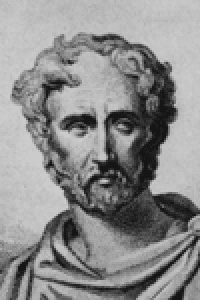
In Roman times Pliny the Elder (WHOSE PHOTO IS AT THE SIDE) compiled four volumes on zoology in his 37-volume treatise called Historia Naturalis. Although widely read during the Middle Ages, they are little more than a collection of folklore, myth, and superstition. One of the more influential figures in the history of physiology, the Greek physician Galen, dissected farm animals, monkeys, and other mammals and described many features accurately, although some were wrongly applied to the human body. His misconceptions, especially with regard to the movement of blood, remained virtually unchanged for hundreds of years. In the 17th century, the English physician William Harvey established the true mechanism of blood circulation.

Until the Middle Ages, zoology was a conglomeration of folklore, superstition, misconception, and descriptions of animals, but during the 12th century it began to emerge as a science. Perhaps the most important naturalist of the era was the German scholar St. Albertus Magnus, who denied many of the superstitions associated with biology and reintroduced the work of Aristotle. The anatomical studies of Leonardo da Vinci were far in advance of the age. His dissections and comparisons of the structure of humans and other animals led him to important conclusions. He noted, for example, that the arrangement of joints and bones in the leg are similar in both horses and humans, thus grasping the concept of homology (the similarity of corresponding parts in different kinds of animals, suggesting a common grouping). The value of his work in anatomy was not recognized in his time. Instead, the Belgian physician Andreas Vesalius is considered the father of anatomy; he circulated his writings and established the principles of comparative anatomy. See ANATOMY
Classification dominated zoology throughout most of the 17th and 18th centuries. The Swedish botanist Carolus Linnaeus developed a system of nomenclature that is still used today—the binomial system of genus and species —and established taxonomy as a discipline. He followed the work of the English naturalist John Ray in relying upon the form of teeth and toes to differentiate mammals and upon beak shape to classify birds. Another leading systematist of this era was the French biologist Comte Georges Leclerc de Buffon. The study of comparative anatomy was extended by such men as Georges Cuvier, who devised a systematic organization of animals based on specimens sent to him from all over the world.Although the word cell was introduced in the 17th century by the English scientist Robert Hooke, it was not until 1839 that two Germans, Matthias Schleiden and Theodor Schwann, proved that the cell is the common structural unit of living things. The cell concept provided impetus for progress in embryology, founded by the Russian scientist Karl von Baer, and for the development by a Frenchman, Claude Bernard, of the study of animal physiology, including the concept of homeostasis (the stability of the body’s internal environment). See CELL,; PHYSIOLOGY,.The organization of scientific expeditions in the 18th and 19th centuries gave trained observers the opportunity to study plant and animal life throughout the world. The most famous expedition was the voyage of the Beagle in the early 1830s. During this voyage, Charles Darwin observed the plant and animal life of South America and Australia and developed his theory of evolution by natural selection. Although Darwin recognized the importance of heredity in understanding the evolutionary process, he was unaware of the work of a contemporary, the Austrian monk Gregor Mendel, who first formulated the concept of particulate hereditary factors—later called genes. Mendel’s work remained obscure
Activity:
Read the scripture on the history of zoology.
trace the history of zoology by making a concept map.
Reference: http://www.history.com/encyclopedia.do?articleId=226469
Thursday, May 28, 2009
Branches of Zoology
Study on the branches of zoology and its field.
Click here for more information about zoology.
Some of the branches of zoology
Determine, which of the following illustrations can be matched to the following studies
1. Herpitology
2. Ornithology
3. Ichthyology
4. Cetology
5. Equinology
6. Myrmecology
7. Mammalogy
8. Primatology
9. Mellitology
10. Apiology
11. Ethology
12. Animal Nutrition
13.Malacology
13. Conchology
14 Cynology
15. acarology
Click here for more information about zoology.
Some of the branches of zoology
Determine, which of the following illustrations can be matched to the following studies
1. Herpitology

2. Ornithology

3. Ichthyology

4. Cetology

5. Equinology

6. Myrmecology

7. Mammalogy

8. Primatology

9. Mellitology

10. Apiology

11. Ethology

12. Animal Nutrition

13.Malacology

13. Conchology

14 Cynology

15. acarology

Tuesday, May 19, 2009
Monday, May 18, 2009
The activity
Here is an activity for you to do while at home.
Click here
Click here for some information about your heart
Click here
Click here for some information about your heart
Sunday, May 17, 2009
The Heart
I. Learning Objectives
General Objective:
Describe the anatomy and physiology of humans.
Specific Objectives:
At the end of the 60 minutes period, 85% of the students are expected to:
1. describe the parts and functions of the human heart through:
1.1 viewing a video clip,
1.2 reading information given in an article or hand outs,
2. appreciate the role played by the heart in the human body,
3. dramatizes how the heart pumps the blood, how each of the structures work.
II. Topic: The Human Heart
III. Resources
A. Materials:
For teaching/ instruction
1, Text and reference books
2. Stop watch
For students
1. Computer(Desktop or Laptop)
2. Heart model/real heart (heart of a pig)
3. Hand outs/article
B. References:
Marieb E.N., Essentials of Human Anatomy and Physiology 7th edition.
The Human Heart http://www.worldinvisible.com/apologet/humbody/heart.htm
How the Body Works (The Heart) http://kidshealth.org/kid/htbw/_bfs_CSactivity.htm
IV. Introduction:
 The heart is one of the most important organs in the entire human body. It is really nothing more than a pump, composed of muscle which pumps blood throughout the body, beating approximately 72 times per minute of our lives. The heart pumps the blood, which carries all the vital materials which help our bodies function and removes the waste products that we do not need. The human heart has 4 chambers. There have been questions concerning how the heart functions, how it looks likes, what are the structures that can be found in the heart, and how these structures works. In order for these questions to be addressed, there will be stations that will set up for the students to choose from. After doing their tasks in their stations, there will be inputs given in each stations concerning; the human heart, its structures, and the functions of each the structures.
V. Learning Experiences/Learning Tasks
I. Activity
There will be two stations that the students will go through during the activity. The following stations are:
1. Information technology station
• They will view the video clip on circulatory system.
The Human Heart
2. Reading and language station
They will be given an article regarding heart.
They will read an article regarding heart.
Click here for you to read an article about heart
In their stations, they will have to manipulate and follow the directions and instruction given to them. They are only given 3 minutes in each station. After three minutes they have to move to another station. There will be guide questions to be given, on the given guide questions, they are going to discuss their answers. The groups are given 3-5 minutes to have their brainstorming, discussion etc regarding their answers.
After synthesizing the answers of the students, the students will then be given another task for them to do. The students will then, dramatize the circulation of blood by showing how the heart pumps blood and how each of the structures work. The class is only given 3-5 minutes to conceptualize their dramatization that will only last for 2-3 minutes. Each members of the group must have their role. Somebody must act as the lungs, act as blood, act as the chambers etc.
II. Analysis
I. There will be questions that will be given to the learners before the lesson proper begins.
Have you been in love?
What part of your body that will react if you will see the person that you love?
Do you have a heart?
What is a heart?
Are you familiar with the parts and structures of your heart?
Do you know how these structures work?
II. Sample Questions for to be asked to the students in their stations.
What is the main function of the heart?
If you are to describe the function of a heart in one word, what would it be? Why?
For you, how important your heart is?
What are the different structures found in a heart?
How these structures work?
If you are to describe the functions of each of the structures, what would it be? Why?
What do you think are the problems that may hinder the functioning of your heart?
What do you think will happen if one of the structures of your heart will not function? Give 1 structure and explain why?
If you are to judge the value of the heart in terms of its structures and functions, how would it be?
How do you feel when you read the article, when you browsed the presentation/watch the video clip and when you manipulated the model of a heart together with your group mates?
III. Abstract
The discussion of the students’ answers will be done. The synthesis of the students’ answers will be done by the teacher.
IV. Application
1. Dramatization of the circulation of blood will be done by each of the students. The group will be given 3-5 minutes to decide on the flow of their dramatization. They have to distribute the role of the members of the group.
2. The students will be given an activity sheet; they will label the parts and structures of the human heart and give its functions.
3. In your Daily experiences, how are you going to apply the concepts presented in the lesson?
V. Agreement
Conduct an interview to a person who is suffering any heart problem.
General Objective:
Describe the anatomy and physiology of humans.
Specific Objectives:
At the end of the 60 minutes period, 85% of the students are expected to:
1. describe the parts and functions of the human heart through:
1.1 viewing a video clip,
1.2 reading information given in an article or hand outs,
2. appreciate the role played by the heart in the human body,
3. dramatizes how the heart pumps the blood, how each of the structures work.
II. Topic: The Human Heart
III. Resources
A. Materials:
For teaching/ instruction
1, Text and reference books
2. Stop watch
For students
1. Computer(Desktop or Laptop)
2. Heart model/real heart (heart of a pig)
3. Hand outs/article
B. References:
Marieb E.N., Essentials of Human Anatomy and Physiology 7th edition.
The Human Heart http://www.worldinvisible.com/apologet/humbody/heart.htm
How the Body Works (The Heart) http://kidshealth.org/kid/htbw/_bfs_CSactivity.htm
IV. Introduction:
 The heart is one of the most important organs in the entire human body. It is really nothing more than a pump, composed of muscle which pumps blood throughout the body, beating approximately 72 times per minute of our lives. The heart pumps the blood, which carries all the vital materials which help our bodies function and removes the waste products that we do not need. The human heart has 4 chambers. There have been questions concerning how the heart functions, how it looks likes, what are the structures that can be found in the heart, and how these structures works. In order for these questions to be addressed, there will be stations that will set up for the students to choose from. After doing their tasks in their stations, there will be inputs given in each stations concerning; the human heart, its structures, and the functions of each the structures.
 V. Learning Experiences/Learning Tasks
I. Activity
There will be two stations that the students will go through during the activity. The following stations are:
1. Information technology station
• They will view the video clip on circulatory system.
The Human Heart
2. Reading and language station
They will be given an article regarding heart.
They will read an article regarding heart.
Click here for you to read an article about heart
In their stations, they will have to manipulate and follow the directions and instruction given to them. They are only given 3 minutes in each station. After three minutes they have to move to another station. There will be guide questions to be given, on the given guide questions, they are going to discuss their answers. The groups are given 3-5 minutes to have their brainstorming, discussion etc regarding their answers.
After synthesizing the answers of the students, the students will then be given another task for them to do. The students will then, dramatize the circulation of blood by showing how the heart pumps blood and how each of the structures work. The class is only given 3-5 minutes to conceptualize their dramatization that will only last for 2-3 minutes. Each members of the group must have their role. Somebody must act as the lungs, act as blood, act as the chambers etc.
II. Analysis
I. There will be questions that will be given to the learners before the lesson proper begins.
Have you been in love?
What part of your body that will react if you will see the person that you love?
Do you have a heart?
What is a heart?
Are you familiar with the parts and structures of your heart?
Do you know how these structures work?
II. Sample Questions for to be asked to the students in their stations.
What is the main function of the heart?
If you are to describe the function of a heart in one word, what would it be? Why?
For you, how important your heart is?
What are the different structures found in a heart?
How these structures work?
If you are to describe the functions of each of the structures, what would it be? Why?
What do you think are the problems that may hinder the functioning of your heart?
What do you think will happen if one of the structures of your heart will not function? Give 1 structure and explain why?
If you are to judge the value of the heart in terms of its structures and functions, how would it be?
How do you feel when you read the article, when you browsed the presentation/watch the video clip and when you manipulated the model of a heart together with your group mates?
III. Abstract
The discussion of the students’ answers will be done. The synthesis of the students’ answers will be done by the teacher.
IV. Application
1. Dramatization of the circulation of blood will be done by each of the students. The group will be given 3-5 minutes to decide on the flow of their dramatization. They have to distribute the role of the members of the group.
2. The students will be given an activity sheet; they will label the parts and structures of the human heart and give its functions.
3. In your Daily experiences, how are you going to apply the concepts presented in the lesson?
V. Agreement
Conduct an interview to a person who is suffering any heart problem.
Saturday, May 16, 2009
Subscribe to:
Posts (Atom)